How Do ELISA Kits Identify Antibodies?
Nov 10th 2022
There are many instances within life sciences where timely antibody detection and quantification within a sample is effective. It can help identify immune responses in vaccinated or infected individuals, detect the protein expression service on the cell's surface, or perform quality control testing. However, you had a quality tool capable of making these detections.
Enzyme-Linked Immunosorbent Assay (ELISA) kits detect multiple antigens and proteins in peptide synthesis service. They are the most common immunoassay type used to detect the presence and measure specific antibodies' concentration in a sample. The ELISA test has proven invaluable as a research and diagnostic tool and plays a crucial role in surveying the proportion of people exposed to infection. This article examines how you can use ELISA kitsto identify antibodies and detect viral infections.
What are ELISA's kits?

The ELISA test is a lab test that measures the concentration of an analyte, which refers to the antibodies or antigens in a particular solution. The immunological assay measures the number of antibodies, such as protein production service and custom antibody production. An ELISA test relies on binding antibodies to their targets to facilitate detection.
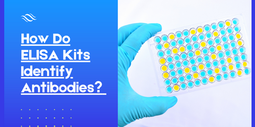
In diagnostics, Cell-based ELISA tests help determine whether and how many antibodies are produced in response to pathogen exposure or vaccination. It usually performs a 96-well plate format, making it easy to screen multiple samples simultaneously. A format that makes it amenable to screening many samples at once. The ELISA test makes it easy to detect quantitative results, which sets it apart from similar trials.
Most ELISA kits detect common sample types such as serum, plasma, and tissue homogenates. Other samples like urine, saliva, cerebral spinal fluid, and other body fluids are also suitable for ELISA kits. The presence of sufficient analyte concentration in the sample is necessary for detection. The samples can comprise buffer components with similar antigenic epitopes. They can also incorporate protease factors that risk affecting assay performance.
Types of ELISA tests

It's essential to consider different ELISA test types. The ELISA test was initially conceptualized in 1971 as researchers wanted a technique to detect antigens and antibodies. This enzyme-based alternative replaced the radioimmunoassay method, which was sometimes dangerous.
There are four ELISA tests for identifying antibodies. While there are different ways of antigen detection, the tests use a similar principle in antibody detection.
Direct ELISA
Direct ELISA test is the fastest ELISA testing type with fewer steps and is less prone to errors. With direct ELISA, antibodies or antigens are absorbed directly into the plate in a nonspecific manner. A conjugated detection antibody then binds to the target protein when applied. You then add a substrate to produce a signal proportional to the amount of analyte in the sample. In this method, a detection substrate changes the sample color quantified on a reader.
The drawbacks of this Direct ELISA type are that it has a non-specific adsorption step and the background noise is too high. The essay requires several steps, which reduces the chances of any errors. The lack of an extra step eliminates the risk of assay sensitivity or amplifying the signal. A secondary antibody step also means no signal amplification, reducing assay sensitivity. This ELISA type also requires conjugated detection antigens/antibodies to be created for each target.
Indirect ELISA
The indirect ELISA test type works similarly to a direct method, though it uses a two-step process in detection. This extra step helps overcome limitations experienced with the direct method and amplifies the test signal.
With the indirect TNFA ELISA, you have a second step that negates using target-specific antibodies for Elisa tests. This is because you will use specific species in the primary antibody without needing secondary antibodies. The indirect test has increased sensitivity compared to the direct ELISA, though it costs less as fewer label antibodies are required.
However, this ELISA type still has some background noise issues because the total sample article binds to the plates. If you use the assay for detecting antibodies, you will need to coat purified target antigens onto the plates. This is the only way to reduce background noises during the tests.
Sandwich ELISA
Sandwich ELISA is a method where two more steps are involved, and the results are precise. It is a common Elisa type where you use two specific antibodies to sandwich an antibody, improving assay sensitivity and specificity. This enzyme-linked type uses indirect TNF Alpha ELISA formats instead of the non-specific antigen binding. It also uses capture and detection antibodies, where the capture antibody adds specificity to the technique, and the detection antibody improves its sensitivity.
However, this sandwich ELISA method requires that you find a capture antibody compatible with the detection antibody to function. A lack of compatibility can affect cross-reactivity during the Elisa tests. Compared to other methods, the sandwich ELISA test has more steps, which creates room for errors. However, since the antigen-binding step is selective, this method is ideal when dealing with complex antigen mix by eliminating the need for purification.
Competitive ELISA
The last ELISA test is competitive or inhibition ELISA, which applies when only one antibody is available or when the analyte is small. The ELISA principle works by detecting interference to a signal level expected from an output, which allows it to produce an inverse.
Competitive ELISA has some advantages and limitations depending on the format you adopted. However, it helps when the antigen is small, as it limits the ability of two antibodies to bind concurrently. When you apply more targets in the sample, there will be a lower output signal. Fortunately, you can achieve multiple formats by adapting the sample to a different format.
The different assay formats all rely on how the ELISA kit manufacturer has designed the binding of the target on the plate surface. You will need to employ a detection step involving a conjugated antigen or antibodies to enable successful binding to be detected and quantified. In most cases, you use colorimetric detection in ELISA testing.
How does an ELISA kit work?

In most cases, the ELISA kits come with easy-to-follow protocols and detailed instructions if you have limited familiarity with them. Since each type of Elisa has different protocol variations, here are the stages to follow in determining how ELISA works.
Plate coating
The basic ELISA test procedurestarts with a coating step where you cover a polystyrene plate with a solution that has antibodies or antigens. The step involves immobilizing an antigen present in the sample to the wells of a microtiter ELISA plate using a passive adsorption approach. In most cases, the results of this plate coating depend on whether you apply an antigen or an antibody sample. If the sample is antigen-specific, the plate will be antigen-bound. However, if you apply purified antigens or capture antibodies, the result is a plate with particular properties.
Cover these plates with an antigen or inactivated form of the virus. You can do this by adsorption into the plate's surface or using a capture antibody. If the blood contains antibodies, it binds to the antigen, and you add a substrate solution to the well. The reaction will produce a color change that detects antibodies. Several plate types are available with different binding properties depending on your assay. Use treated plates for the coating because it allows covalent linkage.
Incubation
After immobilization, add a detection antibody to the ELISA plates, which binds to the adsorbed antigen. Incubating the plates facilitates reaction and binding depending on the assay type. You will also need to perform the blocking phase, where a protein-based solution covers the unbound sites on the plate.
Washing
This is another essential step in ELISA testing after each assay and detection component. Use a buffer to wash the wells after emptying them, ensuring every residue from the incubation is removed. Failing to wash the wells can cause high background signals, while extreme watching can cause lower sample signals. Consistent washing can also result in predictable outcomes.
Blocking
After coating the TS ELISA plates, blocking ensures that the detection antibodies are not specific to the plate. During the blocking phase, you add mixed proteins to the plate, so they occupy other non-specific binding areas. Ensures the proteins don’t relate to the assay when incubating them. If you don’t block the plates during the Elisa tests, your assay will have reduced sensitivity due to higher background noise.
Detection
The last step involves detection, where an enzyme-conjugated antigen or antibody binds to the target antigen-antibody. The detected antibody is directly conjugated to an enzyme or provides a binding site for a labeled secondary antibody.
Every ELISA PPT passes through this step, ending with a color change to show the presence of antibodies. This is where you apply an antibody into a solution and monitor whether the antibodies bind to an antigen. The intensity of the changes in color signals the number of antigens present in the solution. You apply an enzyme-conjugated antibody to test wells where it will bind if its target is current. When you add an appropriate substitute for the enzyme to the plate, it causes a color change proportional to the amount of target bound inside the well.
If it's an HMGB1 ELISA-based test, do the test to detect the immunoglobulin G antibody. The body produces the immunoglobulin M (IgM) and the immunoglobulin G (ImG) antibodies to fight against pathogens. In the final step, place a substrate in the plate, and the enzyme reaction gives the signal.
What do the ELISA kits test?

The results of ELISA kits can be quantitative, qualitative, or semi-quantitative. Usually, you graph these results to compare them with other results and come to a definitive outcome. The quantitative assay uses a serial dilution of a known standard to generate a standard curve. You can calculate the precise quantity of targets in the unknown sample. In a qualitative assay, you collect data numerically using a microplate reader. You then interpret results as positive or negative by comparing them to blank or negative wells without a standard curve.
ELISA kit tests are one of the most sensitive immunoassay tests available today. The sensitivity of the results depends upon the characteristics of the antibody-antigen reaction. You can improve the ELISA test results by adding a substance like those that gives an enhanced chemiluminescent or fluorescent signal.
You will also need to choose suitable experimental antibodies for the ELISA test, especially when using multiple antibodies. You can use both monoclonal and polyclonal antibodies, each with advantages and limitations. For example, monoclonal antibodies produce high specificity, while polyclonal antibodies can bind a target at multiple binding sites and improve sensitivity.
Uses of ELISA's kits
You can use antibody ELISA tests as a supplementary test for a suspect with RT-PCR negative cases for COVID-19. They can also be supplementary detection indicators in diagnosing suspected COVID-19 cases. You can also use the ELISA test in pregnancy, forensic drugs, cancer biomarkers, biosimilars, and biopharmaceutical testing.
Other uses include the detection of food allergens and infectious disease diagnostics. For example, you use the ELISA test to detect infectious agents and antibody responses induced in the host. A sustainable antibody response can be seen in individuals when the microbe cannot.
The benefits of ELISA tests include high specificity, high sensitivity, high throughput, and convenience. The clinical specificity of ELISA's tests is 96%, while the sensitivity is up to 99%. You can use the ELISA tests to process over 20,000 assays in one day, as they are suitable for high throughput tests.
How ELISA's test for HIV works

Most ELISA tests for HIV are based on the antigen-antibody interaction. On the surface of HIV are a lot of proteins that act as antigens, with the early infection called protein 24 (p24). Antigens for HIV are detectable in people around 16 days after infection, while antibodies take longer to produce, usually detectable until 4-12 weeks after infection.
The best ELISA test for HIV can detect antibodies to determine if someone has antibodies to a particular antigen. They look for antibodies in the blood. During the rapid tests, antigens for HIV are fixed on one strip along the immediate test stick. Place a chemical called buffer at the end of the testing stick to facilitate the testing process. This chemical causes the antibodies in the blood to flow along the test's stick.
When the antibodies pass over the section with the antigens, if there are antibodies for HIV present, then they will stick to these antigens and change color. Once the test is complete, the number of stripes will determine whether there is a positive or negative result. One strip represents a negative impact, two represent a positive result, and no strips mean the test did not work correctly.
The LAM ELISA kits can also detect p24 antigen, a sandwich ELISA method. Add a blood sample to a petri dish with antibodies during the process. If the sample is HIV-positive, the p24 antigen on the surface of HIV will attach to the antibodies, and if the sample is negative, then there will be no antigens on, and the dish remains the same.
Bottom line
ELISA tests are standard tests to determine whether someone has antibodies to a particular antigen. You can use cytokine ELISA kits to detect hundreds of proteins and molecules, including cytokines, growth factors, and markers for infectious diseases. The kits are quick, convenient, and accurate research tools to detect quantitative targets of interest in cultures and samples.

