Custom Antibodies: A Critical Tool For Immunohistochemistry
Dec 21st 2023
Cells are the building blocks of life. This is widespread knowledge. But guess what makes up a cell? Proteins. These chains of amino acids are present in unicellular and multicellular organisms. Helping in several fundamental processes of life. But even more importantly, they bring other vital cellular compounds like antibodies to life.
Since their earliest reference in 1890 by Emil von Behring and ShibasaburaKitasator, antibodies have come a long way. Today, about 100 therapeutic antibodies are in use, with the first clinical application approved in 1986. But one of the most pivotal applications of antibodies, particularly custom antibodies, is diagnostics through antigen identification.
Researchers understanding the specificity of custom antibodies employ them in immunohistochemistry. This guide will explore the vital roles custom-made antibodies play in surmounting the shortcomings of immunohistochemistry as a laboratory test.
Let’s get into it by first explaining immunohistochemistry in layperson’s terms.
What is Immunohistochemistry?

Photo by National Cancer Institute on Unsplash
Immunohistochemistry, shortened as IHC, is a standard laboratory test used by pathologists to find and detect early signs of a particular condition in a tissue sample. Among the conditions that immunohistochemistry can help diagnose are cancer, muscular dystrophy, Parkinson’s disease, and Alzheimer’s disease.
But when you carefully consider the name, you’ll notice it combines three terms. These should quickly clue you about the processes it involves:
- Immuno - your immune system, which notices an antigen by secreting antibodies to locate and neutralize parasites, bacteria, and viruses.
- Histo- Just a fancy word for tissue.
- Chemistry - Alludes to the study of microscopic compounds, including human cells.
How do IHC Tests work?
Since every IHC test consists of three primary ingredients: a target antigen, antibodies, and a tissue sample, understanding each play helps explain how the test works. For starters, the target antigen serves as a marker. This means its presence or absence is indicative of a specific disease, condition, or factor.
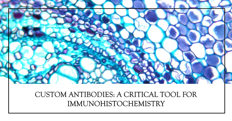
Once your antibody pinpoints the antigen, it will attach to its epitopes. You can liken the binding procedure to how a lock and key works. In this case, the lock is the antigen, and the antibody serves as the key. After binding to a specific antigen, the tissue sample displays a particular stain or color to confirm this lock-key interaction took place. You can observe the staining under a microscope.
Below is a quick breakdown of how typicalimmunohistochemistry techniques work:
- A scientist links an antibody and an enzyme (the enzyme will react upon the binding of the antibody to the antigen of interest).
- The antibody and the antigen of interest bind together, provided the antigen is available.
- This bonding triggers a reaction from the enzyme.
- The enzyme reaction results in staining in the tissue sample, which shows a specific color under a microscope.
For credible outcomes, each step requires meticulous planning and care.
Scenarios Involving the Use of Immunohistochemistry

Photo by National Cancer Institute on Unsplash
Researchers resort to immunohistochemistry applicationsin multiple situations. Some examples are:
- Diagnosing a condition
- Staging and grading prognosis (e.g., cancer)
- Forecasting a patient’s response to treatment
- Track treatment response
- Developing novel drugs
- Identifying disease-causing pathogens (Did you know the first recorded IHC strain was a pneumonia bacteria?).
Now, let’s zoom in on custom antibodies and their roles in successful IHC testing.
Antibodies in Immunohistochemistry
The entire principle and application of immunohistochemistry relied on monoclonal and polyclonal antibodies. Due to the critical nature of the IHC test in pathology, the selection of antibodies is essential for the success of each case.
Primary Antibody
The onset of IHC testing requires the application of a primary antibody highly specific to the antigen of interest. Polyclonal antibodies display high affinity to several epitopes. So they also bind to them. In case you’re wondering, epitopes are simply specific parts of a target antigen.
However, there’s a downside to this multiple affinity displayed by polyclonal antibodies. They are also more likely to react to non-target antigens.
Shifting your gaze to monoclonal antibodies, you’llobserve affinity to a single epitope on an antigen. Hence, this property of the monoclonal category of antibodies means pathologists who employ them get more specific and cleaner staining.
But if your guess is good, they also have one notable downside. Monoclonal antibodies produce less intense and sensitive stains. Some of these setbacks are why secondary antibodies exist in IHC.
Some considerations to keep in mind when picking a primary antibody include:
- In which species was it developed?
- Does it have an affinity for the target protein?
- Has it proven applicable in your sample before?
Secondary Antibody

Photo by National Cancer Institute on Unsplash
To put it shortly, secondary antibodies react to their primary counterparts by binding to them. This process goes by the name of indirect IHC. You’ll often observe multiple secondaries attaching to one primary to heighten staining intensity.
Recently, scientists have turned to custom monoclonal antibodies and their polyclonal counterparts for solutions to some downsides of monoclonal and polyclonal antibody use in IHC.
Custom Antibodies in IHC
As new proteins emerge in the life sciences, research, and biochemical fields, researchers turn to custom antibody services to identify target proteins. Some of the best services offer different types of antibodies to suit the particular needs of their customers.
Those looking to invest in custom antibodies for their IHC test will usually have the following two choices:
- RTU antibodies
- Concentrate antibodies
RTU vs. Concentrate Antibodies
The two main options you’ll consider when consideringcustom antibody developmentexhibit pretty unique properties that set them apart from each other. Firstly, concentrates are adaptable, have lower prices, and are usually suitable for manual or automated staining systems. Despite their flexibility, their use is subject to manufacturer directions.
Also, the optimal dilution of concentrates is adjustable to balance cost, quality, and staining time. They also allow a more comprehensive range of laboratory practices and protocols. If you like the sound of all these perks, you should also know you’ll have to spend more time to time and validate concentrates. Furthermore, your staining quality could be compromised since these types lack definite properties and stability.
RTUs, on the other hand, offer advantages like:
- Improved laboratory efficiency
- Easily manageable reagent
- Saves time on assay dilution, preparation, and antibody validation
Read-to-use antibodies also have better consistency with fewer run-to-run variations. This is particularly noticeable in cases involving automated stainers and associated detection systems.
Manufacturers typically accompany RTUs with verified expiry and a specific number of tests. Therefore, they help streamline antibody management for IHC. Moreover, RTU custom antibodies can drive laboratory growth since they make adopting new antibody assays easier with their reduced validation efforts.
Custom Antibody Specificity
Nothing demonstrates a custom antibody’s (or any antibody for that matter) specificity like the lack of staining in cells or tissues that have their target protein knocked out. If you’re looking to confirm specificity, this should be your go-to sign.
But that’s not all. Other indicators include:
- No staining in cells or tissues known to lack protein expression.
- Staining patterns that conform to widely accepted localization of the target protein in control tissues or cells.
- Identification of one band in western blotting.
To help illustrate the results attainable with the right custom antibody and their role in immuno-chemistry, let’s look at a case study.
Custom Antibody Enable Discovery Involving IHC
Using the Immunogenicity Algorithm, scientists developed custom antibodies specified to RDA288/PAIRBP1. The entire process involved the identification of five peptide sequences matching the criteria for immunogenicity. But before you start scratching your head, the back story.
Progesterone (P4), a hormone involved in the menstrual cycle and pregnancy, was initially believed to act through receptor proteins in its target cells’ nuclei. The general thought was that the nuclear P4 receptor proteins attached to P4-specific promoter regions in several genes to modify their expression. One example of P4 target cells is the follicular granulosa in the female ovary. P4’s primary impact on these cells is the inhibition of apoptosis, enabling the cells to sustain egg maturation.
Another effect of the rising levels of P4 hormones in the said cell’s nucleus is follicular ovulation, among others. Interestingly, these cells also display a self-stimulating mechanism whereby they synthesize P4.
However, this mechanism presents one dilemma - The timing!
Studies revealed that the P4’s known nuclear receptor proteins (PR-A and PR-B), which are separately processed products of the gene, aren’t yet present in the initial phases of follicular growth. Nevertheless, P4 successfully inhibits apoptosis of granulosa cells at this phase. It turns out PR-A and PR-B gene products only became available after the gonadotropin surge. However, P4 suppression of apoptosis can be observed before the gonadotropin spike.
Thus, it’s safe to assume progesterone through other receptors apart from PR-A and P-B.
Dr. John L. Peluso of the University of Connecticut Health Center, Farmington, CT, discovered reasons to think P4’s effects originated at the plasma membrane of early-stage granulosa cells. For instance, he observed direct binding of fluorescein-labeled P4 and nuclear P4 receptor antibody C-262 on the cell membrane. He proceeded to isolate a 60kDa protein after membrane preparations from the C-262 attached to an affinity column.
Dr. John then used mass spectrometric analysis and limited proteolysis to sequence this protein, which he called “RDA288”. And this is where the custom antibody enters the fray.
Custom Antibodies Specific to RDA288/PAIRBP1
At this point, Dr. Peluso sought acustom antibody manufacturer to develop antibodies specific to this novel protein. After developing the five peptide sequence antibody earlier mentioned, Dr.Peluso picked the one nearest the N-terminus of the protein.
Then, the manufacturers added a synthetic spacer amino acid and cysteine to the sequence’s N-terminus, producing the 12-mer peptide ([CZ KQL RKE SQK DRK N). Using a keyhole limpet hemocyanin (KLH- conjugate, the manufacturer injected two laying hens in the breast for about seven weeks. After the fourth injection, they collected the eggs from these layers for three weeks.
They then purified the IgY fraction from 12 eggs and later isolated affinity-purified antibodies from them.
These isolated custom antibodies helped Dr. Peluso to better understand this new membrane-localized, P4-related signaling pathway. In his first research paper, Peluso indicated that the custom antibodies identified a mono-band at the previously assumed molecular weight via western blot analysis and immunocytochemistry. It was also located at the predicted subcellular spot - the plasma membrane.

Photo by Trnava University on Unsplash
Even more interesting, the doctor discovered these custom chicken IgY antibodies blocked P4’s inhibitive apoptosis properties when they reacted with RDA288/PAIRBP1. However, Dr. John Peluso later realized RDA288/PAIRBP1 wasn’t the primary P4 receptor but interfaced with another protein called Progesterone Receptor Membrane Component 1 (PGRMC1). This protein-protein reaction was a prerequisite for P4 to obstruct granular cell apoptosis.
This custom antibody was instrumental because it worked for mice, rats, and humans and is applicable not only to immunohistochemistry but also to immunocytochemistry involving paraffin sections, IP protocols, and Western blots. Lastly, it could also serve as a blocking antibody.
The Role of Custom Antibody Services
The case above shows there might not be readily available custom antibodies on the market in your case. But no need to worry. Custom antibody services exist to meet the challenges presented by cutting-edge research and diagnostic studies. Seeing the rising demand for reliable and verifiable recombinant antibodies, these manufacturers devote time and attention to producing the antibodies you need. You can contact a supplier for custom polyclonal antibodiesor immunohistochemistry antibodies if you need:
- Vital bioreagents for your diagnostic assays
- Research-use-only (RUO) and low-cost antibodies
- Therapeutic-viable recombinant peptides
Antibody experts at companies like Biomatik have several antibody discovery platforms to accommodate the unique requirements of clients. From protein expression services to antigen design and commercial protein services, you can meet all your needs with the right supplier.
Ready to Expand Your Research Horizons with Custom Antibodies?
Custom antibodies are treasured assets in the research, medical, and diagnostic world for their binding affinity, selectivity, and specificity. When you combine these qualities with their animal-friendly production procedure, recombinant systems, and blocking ability, custom antibodies can become indispensable tools in immunohistochemistry. Presently, they have several diagnostic uses reaching far beyond the boundaries of immunoassay applications.
However, you’ll need a tried and tested supplier to fully exploit the potential of antibody applications.
Fortunately, Biomatik offers an unparalleled range of recombinant antibodies, IHC antibodies, and peptide panels crucial for several laboratory tests, including immunohistochemistry. We also supply ELISA kits, testing reagents, and accessories expertly designed to enable our customers to obtain sensitive and reproducible outcomes.

